
2D icons › Biochemistry
Biochemistry: 140 flat 2D icons
Flat vector icons drawn by BioDiorama, free to use with no credit required. Every colour is a setting, many have further settings, and they export as real vector in layered SVG, PDF and editable PowerPoint. Open the 2D Studio and type the name into the search box.
Enzyme kinetics · Macromolecular assemblies · Metabolism · Bonding & folding · Structural biology methods · Protein biochemistry techniques · Food carbohydrates · Food proteins · Food fats & oils · Micronutrients
Enzyme kinetics 5
 Enzyme saturation (low to high substrate)
 Concerted (MWC) model of allostery
 Sequential (KNF) model of allostery
 Conformational selection
 Induced fit vs conformational selection
Macromolecular assemblies 15
 Bacterial and eukaryotic ribosomes (70S and 80S)
 Ribosome exit tunnel with nascent chain
 Proteasome ring (top view, seven subunits)
 TRiC/CCT chaperonin (closed, eukaryotic)
 Chaperonin folding cycle (GroEL–GroES)
 ATP synthase (rotary F₀F₁ motor)
 ATP synthase c-ring (top view)
 Viral capsid self-assembly
 Amyloid aggregation pathway
 Microtubule assembly (dimer to tube)
 Actin nucleation and elongation
 Pyruvate dehydrogenase complex
 Fatty acid synthase (FAS dimer)
 Hsp104/ClpB disaggregase threading a protein
 Carboxysome (bacterial microcompartment)
Metabolism 24
 Citric acid (Krebs) cycle
 Glycolysis as an energy staircase
 Glycolysis and gluconeogenesis (bypass steps)
 Urea cycle
 β-Oxidation spiral
 Pentose phosphate pathway
 Electron transport chain (complexes I–IV)
 ATP–ADP cycle
 Malate–aspartate shuttle
 Glycerol-3-phosphate shuttle
 Glycogen particle (branched tree)
 Lipolysis at a lipid droplet
 Lactic acid fermentation
 Alcoholic fermentation (yeast)
 Catabolism overview (food to ATP)
 Proton motive force (gradient across a membrane)
 Cholesterol synthesis (mevalonate pathway)
 Stable isotope tracing (¹³C-glucose)
 Reactive oxygen species detoxification (SOD and catalase)
 Glutathione redox cycle (GSH/GSSG)
 Anabolism and catabolism
 Metabolic network map
 Glucose split into two pyruvates (carbon count)
 Substrate channelling through an enzyme tunnel
Bonding & folding 16
 Hydrogen bond (donor and acceptor)
 Ionic attraction (salt bridge)
 Hydrophobic effect (oil droplets coalescing)
 Van der Waals contact (induced dipoles)
 Disulfide bridges stapling a folded chain
 π–π stacking of aromatic rings
 Cation–π interaction
 Metal coordination (zinc site)
 Protein hydration shell
 Helical wheel (amphipathic helix)
 Protein denaturation by heat
 Refolding after denaturant removal (Anfinsen)
 Salting out a protein (ammonium sulfate)
 Protein–ligand binding equilibrium (Kd)
 Electrostatic surface of a protein (Bonding & folding)
 Hydrophobic collapse of a chain
Structural biology methods 11
 Hanging-drop crystallisation (vapour diffusion)
 Protein crystal in a cryo-loop
 Electron density map with a fitted chain
 Crystal lattice and unit cell
 Bragg's law (diffraction from lattice planes)
 Synchrotron light source (beamlines)
 Cryo-EM grid (holey carbon)
 Plunge freezing (vitrification)
 Cryo-EM 3D density map
 Cryo-electron tomography (tilt series)
 NMR tube (Structural biology methods)
Protein biochemistry techniques 13
 Dialysis bag in buffer
 Dialysis cassette with float
 Protein purification column with fractions
 Elution profile (A280 with salt gradient)
 Size-exclusion chromatography (porous beads) (Protein biochemistry techniques)
 Ion-exchange chromatography (charged beads)
 SDS unfolding and coating a protein
 SDS-PAGE of a purification (lysate to eluate)
 Bradford assay (colour change with protein)
 Centrifugal concentrator (ultrafiltration)
 Isoelectric focusing strip (pI)
 Affinity tag removal by protease
 Coupled enzyme assay (NADH at 340 nm)
Food carbohydrates 16
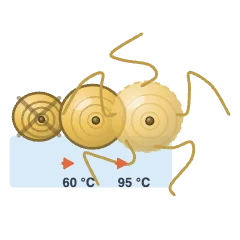 Starch gelatinisation
 Starch retrogradation (staling)
 Amylose (linear α-1,4 helix)
 Amylopectin (branched cluster)
 Starch granule lamellae
 Resistant starch
 Soluble fibre (viscous gel)
 Insoluble fibre (bran particle)
 Pectin gel (junction zones)
 Sugar: crystal, syrup, glass
 State diagram and Tg
 Sucrose inversion
 Maillard reaction
 Caramelisation
 Acrylamide formation
 Hydrocolloid flow behaviour
Food proteins 12
 Gluten network
 Disulfide crosslinking
 Heat-set protein gels
 Protein unfolding at an interface
 Casein micelle
 Rennet coagulation
 Acid coagulation (pH 4.6)
 Whey proteins on heating
 Egg white coagulation
 Collagen to gelatin
 Protein foam and its drainage
 Extrusion texturisation
Food fats & oils 11
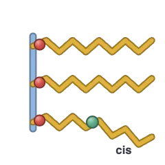 Triacylglycerol
 Fat crystal polymorphs
 Fat crystal network
 Chocolate tempering
 Fat bloom on chocolate
 Oxidative rancidity
 Hydrolytic rancidity
 Antioxidant stopping a chain
 Emulsion droplet interface
 How an emulsion breaks
 Emulsifier HLB
Micronutrients 17
 Vitamin A in vision
 Vitamin D from UV
 Vitamin C as a reductant
 Vitamin E in a membrane
 Vitamin K carboxylation
 B vitamins as cofactors
 Folate and the neural tube
 B12 and intrinsic factor
 Haem and non-haem iron
 What helps and blocks iron
 Calcium and oxalate
 Iodine and thyroid hormone
 Zinc bound by phytate
 Selenium in glutathione peroxidase
 Fortification of a flour stream
 In vitro digestion and bioaccessibility
 Collagen without vitamin C