

2D icons › Cancer
Cancer: 231 flat 2D icons
Flat vector icons drawn by BioDiorama, free to use with no credit required. Every colour is a setting, many have further settings, and they export as real vector in layered SVG, PDF and editable PowerPoint. Open the 2D Studio and type the name into the search box.
Cancer cells · Cancer cell states · Tumours · Tumour in tissue · Genome & evolution · Cell states & plasticity · Metastatic cascade · Metastatic disease · Tumour microenvironment · Hypoxia & metabolism · Angiogenesis & vessels · Surgery & ablation · Radiotherapy · Chemotherapy actions · Targeted therapy · Cell & immune therapy · Resistance · Side effects · Breast & gynaecological tumours · Digestive tumours · Thoracic, urological & endocrine tumours · Skin, brain, bone & childhood tumours · Blood cancers · Mouse tumour models · Genetic & carcinogen mouse models · Other tumour models · Measuring tumours · Screening & diagnosis · Staging & grading · Causes & risk
Cancer cells 18
 Cancer cell (small, round)
 Cancer cell (large, irregular)
 Cancer cell (spiky, with protrusions)
 Carcinoma cell (polygonal epithelial)
 Spindle cancer cell (sarcoma)
 Giant multinucleated tumour cell
 Signet ring cell
 Reed–Sternberg cell
 Leukaemic blast with Auer rods
 Cancer stem cell
 Hairy cell (hairy cell leukaemia)
 Clear cell carcinoma cells
 Small cell carcinoma (oat cells)
 Melanoma cell
 Glioblastoma cell
 Koilocyte (HPV-infected cell)
 Abnormal tripolar mitosis
 Cancer cell with organelles
Cancer cell states 12
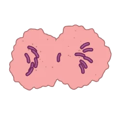 Dividing cancer cell
 Cancer cell dying by apoptosis
 Tumour necrosis
 Dormant cancer cell
 Invading cancer cell (squeezing through the matrix)
 Drug-resistant cancer cell (efflux pumps)
 Hypoxic cancer cell (HIF-1α)
 Therapy-induced senescent cancer cell
 Warburg effect (aerobic glycolysis)
 Cancer cell with micronuclei (chromosomal instability)
 Cancer cell overexpressing a receptor (HER2)
 Cancer cell releasing exosomes
Tumours 8
 Cancer cell cluster (mixed sizes)
 Tumour spheroid
 Tumour spheroid (section): necrotic core
 Solid tumour with blood vessels
 Tumour angiogenesis (VEGF)
 Tumour growth
 Tumour heterogeneity (clones)
 Clonal evolution
Tumour in tissue 11
 Normal epithelium
 Hyperplasia
 Dysplasia
 Carcinoma in situ
 Invasive carcinoma
 Cancer progression (normal to invasive)
 Benign tumour (encapsulated)
 Malignant tumour (invasive)
 Colon polyp (adenoma)
 Polyp to carcinoma sequence
 Squamous cell carcinoma (keratin pearl)
Genome & evolution 6
 Hallmarks of cancer wheel
 Two-hit inactivation of a tumour suppressor
 Tumour phylogenetic tree (truncal and branch mutations)
 Chromothripsis (shattered and rejoined chromosome)
 Microsatellite instability (shifted repeat peaks)
 Oncoprint of recurrent alterations
Cell states & plasticity 11
 EMT spectrum (epithelial, hybrid, mesenchymal)
 Mesenchymal–epithelial transition at a metastatic site
 Cancer stem cell hierarchy
 Perivascular cancer stem cell niche
 Collective invasion led by leader cells
 Amoeboid cancer cell migration through collagen
 Tumour budding at the invasive front
 Entosis (cell-in-cell)
 Drug-tolerant persister cells
 Anoikis resistance (survival after detachment)
 Cancer cell–macrophage fusion hybrid
Metastatic cascade 9
 Cancer cell breaching the basement membrane
 Invadopodium degrading matrix (MT1-MMP)
 Intravasation at a TMEM doorway
 Circulating tumour cell cloaked by platelets
 Circulating tumour cell cluster with a neutrophil
 Tumour cell arrested in a capillary
 Tumour cell extravasation
 Metastatic colonisation of the lung
 Pre-metastatic niche
Metastatic disease 4
 Lymphatic spread to a sentinel lymph node
 Osteolytic bone metastasis (vicious cycle)
 Perineural invasion
 Lymphovascular invasion (tumour embolus)
Tumour microenvironment 17
 Tumour microenvironment (cell types)
 Myofibroblastic CAF (myCAF) wrapping a tumour nest
 Inflammatory CAF (iCAF) secreting IL-6
 Antigen-presenting CAF (apCAF) with MHC II
 TAM–tumour cell EGF/CSF-1 paracrine loop
 MDSCs suppressing a T cell
 Treg depriving an effector T cell of IL-2
 Tertiary lymphoid structure in a tumour
 Neutrophil extracellular traps capturing tumour cells
 Normal and tumour vascular networks
 Tumour-associated collagen signature (TACS-3)
 Desmoplastic stroma around pancreatic cancer glands
 Inflamed, excluded and desert tumours
 Cancer-associated adipocytes feeding tumour cells
 Cancer–immunity cycle
 Neuron–glioma synapse
 Fusobacterium in colorectal cancer
Hypoxia & metabolism 8
 Oxygen gradient away from a tumour vessel
 Pseudopalisading necrosis (glioblastoma)
 Acidic tumour microenvironment (lactate and H⁺ export)
 Metabolic symbiosis (lactate shuttle)
 Glutamine addiction (glutaminolysis feeding the TCA cycle)
 Glucose competition between tumour cells and T cells
 Mutant IDH producing the oncometabolite 2-HG
 Cancer-associated thrombosis (tissue factor)
Angiogenesis & vessels 7
 Angiogenic switch
 Vessel co-option by tumour cells
 Vasculogenic mimicry
 Intussusceptive angiogenesis (pillar splitting)
 Vessel normalisation by anti-VEGF therapy
 Glomeruloid microvascular proliferation
 Tumour lymphangiogenesis (VEGF-C)
Surgery & ablation 9
 Resection margin status (R0 and R1)
 Sentinel lymph node biopsy (blue dye and gamma probe)
 Mohs micrographic surgery (layer-by-layer excision)
 Radiofrequency ablation with an umbrella electrode
 Microwave ablation of a lung tumour
 Cryoablation ice ball around a kidney tumour
 High-intensity focused ultrasound (HIFU)
 Radioembolisation with Y-90 microspheres
 Irreversible electroporation (NanoKnife)
Radiotherapy 2
 Proton Bragg peak versus photon depth dose
 Radioligand therapy (¹⁷⁷Lu-PSMA)
Chemotherapy actions 11
 DNA interstrand crosslinks (alkylating agent)
 Platinum adduct bending DNA
 Antimetabolite stalling DNA synthesis
 Topoisomerase II trapped on cleaved DNA
 Taxane stabilising microtubules (mitotic arrest)
 Vinca alkaloid blocking microtubule assembly
 Anthracycline intercalating between base pairs
 Log-kill with repeated chemotherapy cycles
 Neutrophil nadir after a chemotherapy cycle
 Immunogenic cell death (calreticulin, ATP, HMGB1)
 Tumour lysis syndrome
Targeted therapy 9
 PARP inhibitor synthetic lethality in BRCA-mutant cells
 CDK4/6 inhibitor holding cells in G1
 Oestrogen receptor blocked in a breast cancer cell
 Antibody–drug conjugate: binding, uptake and payload release
 Antibody-dependent cellular cytotoxicity against a HER2 tumour cell
 Anti-VEGF antibody sequestering VEGF
 BCL-2 inhibitor freeing BAX at the mitochondrion
 Differentiation therapy of APL with ATRA
 Proteasome inhibitor in a myeloma cell
Cell & immune therapy 8
 Tumour-infiltrating lymphocyte (TIL) therapy
 CAR-NK cell
 Dendritic cell vaccine
 Personalised neoantigen mRNA vaccine
 Oncolytic virus bursting a tumour cell
 Anti-PD-1 antibody restoring T-cell killing
 Repolarising tumour-associated macrophages (M2 to M1)
 Autologous stem cell transplant cycle
Resistance 8
 Gatekeeper mutation blocking a kinase inhibitor
 Bypass signalling through MET amplification
 CD19-negative relapse after CAR-T therapy
 Lineage plasticity to neuroendocrine cancer
 Selection of a pre-existing resistant clone
 BRCA reversion mutation restoring repair
 Fibroblast-secreted HGF protecting tumour cells from a drug
 Blood–brain barrier as a drug sanctuary
Side effects 1
 Chemotherapy-induced hair loss (anagen effluvium)
Breast & gynaecological tumours 3
 Invasive ductal carcinoma of the breast (histology)
 Invasive lobular carcinoma (single-file cells)
 Cervical intraepithelial neoplasia grades (CIN 1–3)
Digestive tumours 2
 Barrett's oesophagus with early adenocarcinoma (endoscopy)
 Neuroendocrine tumour (nests, salt-and-pepper chromatin)
Thoracic, urological & endocrine tumours 3
 Lung adenocarcinoma (lepidic growth along alveoli)
 Papillary urothelial carcinoma (bladder fronds)
 Gleason patterns 3, 4 and 5 (prostate cancer)
Skin, brain, bone & childhood tumours 4
 Basal cell carcinoma (peripheral palisading)
 Cutaneous melanoma (asymmetric, irregular border)
 Meningothelial whorls and psammoma bodies (meningioma)
 Medulloblastoma (Homer Wright rosettes)
Blood cancers 4
 Follicular lymphoma (back-to-back follicles)
 Chronic myeloid leukaemia blood film (granulocytes at all stages)
 Acute lymphoblastic leukaemia blood film (lymphoblasts)
 Chronic lymphocytic leukaemia blood film (smudge cells)
Mouse tumour models 12
 Cell-line-derived xenograft (CDX) injection into the flank
 Patient-derived xenograft (PDX) fragment implantation
 Serial PDX passaging (P0 to P2)
 Orthotopic injection into the mammary fat pad
 Orthotopic pancreatic tumour implantation (laparotomy)
 Experimental lung metastasis after tail-vein injection
 Intracardiac injection for bone metastasis
 Intrasplenic injection for liver metastasis
 Excised tumours lined up by treatment group
 Dorsal skinfold window chamber for intravital imaging
 Mouse with malignant ascites
 Bilateral flank tumours (irradiated and distant)
Genetic & carcinogen mouse models 7
 Genetically engineered mouse model (Cre-activated Kras and p53)
 MMTV-PyMT mouse with multiple mammary tumours
 Syngeneic tumour in an immunocompetent mouse
 Humanised mouse (human CD34⁺ cells engrafted in NSG)
 ApcMin intestinal polyps (Swiss roll)
 DMBA/TPA skin papillomas on a shaved back
 AOM/DSS colitis-associated colon tumours
Other tumour models 10
 Zebrafish larval xenograft
 Chick chorioallantoic membrane (CAM) tumour assay
 Drosophila eye-disc tumour (RasV12 scrib clones)
 Patient-derived tumour organoid (PDO)
 Tumour organoid drug screen (response heat map)
 Tumour organoid and T-cell co-culture
 Precision-cut tumour slice culture
 Microfluidic extravasation assay
 Soft agar colony formation (anchorage-independent growth)
 Cell line authentication by STR profiling
Measuring tumours 8
 Tumour volume from length and width (V = L × W² / 2)
 Bioluminescence signal growing over weeks
 Ki-67 immunohistochemistry (proliferation index)
 Blood tube for cell-free DNA (plasma separated)
 Tumour cells shedding ctDNA into the blood
 Cell-free DNA fragment-size profile (167 bp peak)
 Circulating tumour cells captured on an antibody-coated micropost chip
 Serum tumour marker falling and rising (CA-125)
Screening & diagnosis 4
 Pap smear with high-grade squamous lesion (HSIL) cells
 Cervical screening sample (brush and liquid cytology vial)
 Faecal immunochemical test (FIT) kit
 Hereditary breast and ovarian cancer pedigree
Staging & grading 6
 T stage by depth of invasion in the bowel wall
 N stage by number of involved lymph nodes
 Breast tumour size categories (T1, T2, T3)
 Breslow thickness of a melanoma
 Tumour grade by differentiation (G1, G2, G3)
 Cancer stages from I to IV (local to distant)
Causes & risk 9
 Tobacco smoke carcinogens reaching the airway lining
 Ultraviolet light reaching epidermal keratinocytes
 HPV E6 and E7 disabling p53 and Rb
 Correa cascade (gastritis to gastric cancer)
 Chronic hepatitis to cirrhosis to liver cancer
 Asbestos body in lung tissue
 Aflatoxin-producing mould on stored peanuts
 Familial adenomatous polyposis (colon carpeted with polyps)
 Field cancerisation in the oral mucosa