

2D icons › Microbes & viruses
Microbes & viruses: 445 flat 2D icons
Flat vector icons drawn by BioDiorama, free to use with no credit required. Every colour is a setting, many have further settings, and they export as real vector in layered SVG, PDF and editable PowerPoint. Open the 2D Studio and type the name into the search box.
Bacterial shapes · Bacterial structures · Named bacteria · Bacterial biology · Archaea · Viruses · Prions & viroids · Virus life cycle · Fungi · Protists & parasites · Microbiome · Food microbiology & spoilage · Selective & differential media · Biochemical tube tests · Identification tests · Culture & counting · Susceptibility testing · Colony morphology · Stains under the microscope · Bacterial species & forms · Biofilm stages & assays · Bacteriophages · Phage infection & defence · Dimorphic & systemic fungi · Dermatophytes & yeasts · Mould structures · Malaria parasite stages · Toxoplasma & trypanosomes · Protists · Helminth eggs · Helminth larvae & adults · Resistance mechanisms · Resistance spread & evolution · Parasite life cycles · Vectors & hosts
Bacterial shapes 14
 Coccus (round bacterium)
 Diplococci (pairs)
 Streptococci (chain of cocci)
 Tetrad (cocci in fours)
 Sarcina (cube of eight cocci)
 Bacillus (rod-shaped bacterium)
 Diplobacilli (rods in pairs)
 Streptobacilli (chain of rods)
 Coccobacilli
 Vibrios (comma-shaped bacteria)
 Spirillum (rigid spiral)
 Fusiform bacteria (spindle-shaped)
 Mycoplasma (wall-less, pleomorphic)
 Caulobacter (stalked and swarmer cell)
Bacterial structures 21
 Polar flagellum (monotrichous)
 Lophotrichous flagella (polar tuft)
 Amphitrichous flagella (one at each pole)
 Peritrichous flagella (all over)
 Bacterial flagellar motor
 Fimbriae (short pili)
 Sex pilus (F pilus)
 Bacterial capsule
 Endospores (central, subterminal, terminal)
 Endospore structure (core, cortex, coat)
 Binary fission
 Bacterial division (FtsZ ring)
 Magnetotactic bacterium (magnetosome chain)
 Outer membrane vesicles
 Gram-positive cell envelope (Bacterial structures)
 Gram-negative cell envelope (Bacterial structures)
 Mycobacterial cell envelope (Bacterial structures)
 Peptidoglycan mesh
 Type III secretion system (injectisome)
 Multidrug efflux pump (AcrAB–TolC)
 Bacterial cell (labelled cut-away)
Named bacteria 37
 Escherichia coli
 Staphylococcus aureus (clusters)
 Streptococcus pneumoniae (encapsulated diplococci)
 Mycobacterium tuberculosis (cording)
 Helicobacter pylori
 Vibrio cholerae
 Treponema pallidum (syphilis spirochaete)
 Borrelia (Lyme disease spirochaete)
 Leptospira (hooked spirochaete)
 Bacillus anthracis (chains)
 Clostridioides difficile (with spores)
 Pseudomonas aeruginosa
 Salmonella invading a host cell
 Listeria with actin comet tails
 Neisseria (kidney-bean diplococci)
 Chlamydia inclusion (elementary and reticulate bodies)
 Bifidobacterium (branched rods)
 Streptomyces (mycelium with spore chains)
 Corynebacterium (palisades, club-shaped rods)
 Campylobacter (seagull-shaped)
 Yersinia pestis (bipolar staining)
 Cyanobacterium filament with heterocyst (Anabaena)
 Spirulina (Arthrospira) helical filament
 Bdellovibrio attacking a bacterium
 Root nodule bacteroids (Rhizobium)
 Purple sulfur bacterium (sulfur globules)
 Lactococcus (ovoid cocci in chains)
 Enterococcus (oval diplococci)
 Klebsiella pneumoniae (encapsulated rods)
 Acinetobacter baumannii (coccobacilli in pairs)
 Bacteroides (pleomorphic gut anaerobe)
 Akkermansia muciniphila in the mucus layer
 Faecalibacterium prausnitzii (butyrate producer)
 Cutibacterium acnes (club-shaped rods)
 Rickettsia free in the host cytoplasm
 Legionella in its ER-studded vacuole
 Wolbachia in an insect cell
Bacterial biology 19
 Biofilm formation (attachment to dispersal)
 Mature biofilm (towers in matrix)
 Bacterial colony on agar (close-up)
 Streak plate (isolated colonies)
 Antibiotic disk diffusion (zones of inhibition)
 Quorum sensing
 Antibiotic lysing a bacterium
 Antibiotic targets in a bacterium
 Antibiotic resistance mechanisms
 Bacterial conjugation
 Bacterial transformation (DNA uptake)
 Transduction (phage carrying bacterial DNA)
 L-form bacteria (wall-deficient)
 Sporulation stages
 Chemotaxis (run and tumble)
 Gut microbiome (Bacterial biology)
 Cholera toxin (AB5) binding a cell
 Lipopolysaccharide (LPS, endotoxin)
 Zipper invasion (Listeria entering a cell)
Archaea 4
 Archaeon with S-layer
 S-layer lattice (top view)
 Square archaea (Haloquadratum)
 Sulfolobus (lobed archaeon)
Viruses 44
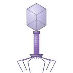 Bacteriophage T4
 Bacteriophage lambda
 Bacteriophage T7 (short tail)
 Filamentous phage M13
 Non-enveloped icosahedral virus
 Enveloped virus (spherical)
 Tobacco mosaic virus (helical rod)
 SARS-CoV-2 (coronavirus)
 SARS-CoV-2 (cut-away with genome)
 Coronavirus spike protein (trimer)
 Influenza virus (HA and NA)
 Influenza virus (cut-away, 8 RNPs)
 HIV (cone-shaped capsid)
 Immature HIV particle (Gag lattice)
 Herpesvirus
 Adenovirus (with fibres)
 Human papillomavirus (HPV)
 Hepatitis B virus (Dane particle)
 Rotavirus (wheel-like triple-layered capsid)
 Norovirus (cup-like surface)
 Poliovirus (picornavirus)
 Rabies virus (bullet-shaped)
 Ebola virus (filament)
 Measles virus (paramyxovirus)
 Zika and dengue virus (flavivirus)
 Poxvirus (brick-shaped)
 Giant virus (mimivirus)
 Adeno-associated virus (AAV)
 Lentiviral vector particle (VSV-G pseudotyped)
 Virus-like particle (empty capsid)
 Capsid cut-away with genome
 Pandoravirus (amphora-shaped giant virus)
 Lemon-shaped archaeal virus
 Geminivirus (twinned particles)
 Baculovirus in an occlusion body
 Respiratory syncytial virus (pleomorphic)
 Hantavirus (surface lattice of Gn–Gc spikes)
 Arenavirus (Lassa, sandy ribosomes inside)
 Influenza haemagglutinin (HA trimer)
 Influenza neuraminidase (NA tetramer)
 SARS-CoV-2 spike binding ACE2
 Antigenic drift (point mutations escape antibodies)
 Antigenic shift (reassortment of segments)
 Herpesvirus latency in a sensory neuron
Prions & viroids 2
 Prion amyloid fibrils (PrPSc)
 Viroid (circular RNA rod)
Virus life cycle 13
 Virus attachment to a receptor
 Virus entry by endocytosis
 Virus–cell membrane fusion
 Uncoating (genome release)
 Viral genome replication
 Virus assembly
 Virus budding
 Cell lysis releasing virions
 Virus life cycle (overview)
 Phage lytic cycle
 Phage lysogenic cycle (prophage)
 Phage injecting its DNA
 Retroviral integration (provirus)
Fungi 23
 Budding yeast (Saccharomyces cerevisiae)
 Fission yeast (Schizosaccharomyces pombe)
 Candida albicans with pseudohyphae
 Candida yeast forming a germ tube (hypha)
 Septate hyphae
 Mycelium (hyphal network)
 Aspergillus conidiophore
 Penicillium conidiophore (brush)
 Rhizopus (bread mould) sporangium
 Fungal spores (conidia)
 Spore germinating
 Basidium with basidiospores
 Ascus with eight ascospores
 Mould colony on agar
 Cryptococcus with capsule (India ink)
 Pneumocystis cysts
 Chytrid fungus (Batrachochytrium)
 Fusarium macroconidia (banana-shaped)
 Alternaria conidia (muriform chain)
 Microsporidian spore (polar tube)
 Yeast mating projections (shmoo)
 Fungal cell wall layers
 Malassezia (bottle-shaped yeast)
Protists & parasites 15
 Plasmodium ring form (early trophozoite)
 Plasmodium schizont (merozoites in a red cell)
 Plasmodium gametocyte (banana form)
 Plasmodium sporozoites
 Toxoplasma tachyzoite (apical complex)
 Trypanosoma (trypomastigote)
 Leishmania promastigote
 Leishmania amastigotes in a macrophage
 Giardia trophozoite
 Entamoeba histolytica trophozoite
 Cryptosporidium oocyst
 Trichomonas vaginalis
 Babesia in red cells (Maltese cross)
 Naegleria (amoeboflagellate)
 Acanthamoeba trophozoite and cyst
Microbiome 7
 Healthy microbiota versus dysbiosis
 Faecal microbiota transplant (capsule)
 Fibre fermented to short-chain fatty acids
 Skin microbiome in a hair follicle
 Vaginal epithelium with lactobacilli
 Gut–brain axis
 Colonisation resistance (commensals exclude a pathogen)
Food microbiology & spoilage 14
 The temperature danger zone
 Water activity and who grows
 pH limits and acid foods
 Hurdle technology
 A spore surviving the heat
 The botulinum cook (F₀)
 Biofilm in a dead leg
 Clean-in-place cycle
 Osmophilic spoilage yeast
 Xerophilic mould on a dry food
 Mycotoxin in a kernel
 Enzymatic browning
 Histamine in fish
 Accelerated shelf-life testing
Selective & differential media 15
 Chocolate agar with Haemophilus colonies
 Mannitol salt agar (yellow haloes round S. aureus)
 XLD agar (black-centred Salmonella colonies)
 TCBS agar (yellow Vibrio cholerae colonies)
 EMB agar (green metallic sheen of E. coli)
 CLED agar urine culture (calibrated-loop streak)
 Chromogenic urinary agar (colour-coded colonies)
 Chromogenic Candida agar (three isolates)
 Sabouraud dextrose agar with mould and yeast
 Löwenstein–Jensen slant (rough buff TB colonies)
 CIN agar (bull’s-eye Yersinia colonies)
 DNase agar (clear zones round positive spots)
 Starch hydrolysis plate (iodine flood)
 Charcoal agar with Bordetella (mercury-drop colonies)
 Bile esculin agar (black haloes of enterococci)
Biochemical tube tests 9
 Triple sugar iron slants (K/K, K/A, A/A, H₂S)
 Simmons citrate slants (green negative, blue positive)
 Christensen urea slants (slow and rapid urease)
 SIM tubes (non-motile stab and motile spread)
 Indole test (red Kovács ring)
 Sugar fermentation with Durham tubes (acid and gas)
 Oxidation–fermentation (OF) glucose tubes
 Gelatin liquefaction (tilted tubes)
 Stormy fermentation in litmus milk
Identification tests 12
 Oxidase test on filter paper
 Slide coagulase test (clumping factor)
 Latex agglutination card (streptococcal grouping)
 Optochin disc on blood agar (pneumococcus)
 CAMP test (arrowhead haemolysis)
 Satellitism round a Staphylococcus streak
 X and V factor discs (Haemophilus growth needs)
 String test (hypermucoviscous Klebsiella)
 MALDI target plate (spotted colonies and matrix)
 Automated identification card with transfer tube
 Multi-compartment identification tube
 Rapid urease test on a gastric biopsy
Culture & counting 19
 Spread plate on a turntable
 Pour plate (lens-shaped colonies in the agar)
 Ten-fold serial dilution and plating
 Counting colonies on a gridded counter
 Drop plate count (Miles and Misra)
 Replica plating with a velvet block
 McFarland turbidity standards against a lined card
 Anaerobic jar with gas-generating sachet
 Candle jar (raised CO₂ for capnophiles)
 Sealed pouch for microaerophilic culture
 Transport swab in charcoal Amies medium
 Urine dipslide and its container
 Membrane filter with coliform colonies (gridded)
 Hanging drop on a depression slide
 Heat-fixing a smear over a Bunsen flame
 Pouring agar plates
 Growth patterns in broth (turbid, sediment, pellicle, flocculent)
 Oxygen requirements in thioglycollate broth
 Flaming an inoculating loop
Susceptibility testing 8
 D-test for inducible clindamycin resistance
 Double-disc synergy test for ESBL (keyhole zones)
 Carbapenem inactivation method (CIM)
 Checkerboard synergy plate (two drugs)
 Agar dilution MIC (spotted plates)
 Antibiotic disc dispenser on a plate
 Resistant colonies growing inside an inhibition zone
 Antibiotic gradient plate (wedge layers)
Colony morphology 15
 Colony forms (circular to filamentous)
 Colony elevations (side profiles)
 Colony margins (entire to curled)
 Mucoid Klebsiella colonies (MacConkey)
 Pigmented colonies (Serratia, Chromobacterium, Micrococcus)
 Pseudomonas with diffusible green pigment
 Fluorescent Pseudomonas colonies under UV light
 Proteus swarming waves on blood agar
 Wrinkled Bacillus subtilis colony (biofilm)
 Chalky Streptomyces colonies with exudate droplets
 Medusa-head colony edge (Bacillus anthracis)
 Draughtsman colonies of pneumococcus (close-up)
 Small-colony variants of Staphylococcus aureus
 Fried-egg Mycoplasma colonies (low power)
 Bioluminescent Vibrio fischeri colonies in the dark
Stains under the microscope 3
 Calcofluor white (fluorescent fungal walls)
 KOH mount of a skin scraping (hyphae over keratinocytes)
 Clue cell beside a normal squamous cell
Bacterial species & forms 13
 Clostridium tetani (drumstick spores)
 Clostridium perfringens (boxcar rods)
 Nocardia (beaded branching filaments)
 Hyphomicrobium budding at the tip of a hypha
 Myxococcus fruiting body (cells streaming into a mound)
 Thiomargarita (chain of giant sulphur pearls)
 Epulopiscium (giant cell with internal offspring)
 Lactobacillus (long rods in chains)
 Filamentous E. coli (division blocked)
 Rod to spheroplast after cell-wall damage
 Spirochaete periplasmic flagella (side and cross-section)
 Prochlorococcus (thylakoids round the rim)
 Anammox bacterium with its anammoxosome
Biofilm stages & assays 12
 Biofilm step 1: reversible attachment
 Biofilm step 2: irreversible attachment
 Biofilm step 3: microcolony in its matrix
 Oxygen and nutrient gradients through a biofilm
 Biofilm step 5: dispersal from a hollowed colony
 Biofilm matrix close-up (polysaccharide, eDNA, amyloid)
 Biofilm lining a catheter (cross-section)
 Crystal violet biofilm assay (stained wells)
 Peg-lid biofilm device (MBEC)
 Flow-cell biofilm under a confocal objective
 Pellicle biofilm at the air–liquid interface
 Persister cells surviving antibiotic in a biofilm
Bacteriophages 8
 Bacteriophage φX174 (spiked icosahedron)
 Bacteriophage φ29 (prolate head, appendages)
 Bacteriophage PRD1 (inner membrane, tube extended)
 Bacteriophage φ6 (enveloped, segmented dsRNA)
 RNA phages (MS2) bound along an F pilus
 Phage DNA packaging motor (portal and terminase)
 Phage capsid maturation (round prohead to angular head)
 Tailocin (R-type pyocin) before and after firing
Phage infection & defence 10
 Plaque types: clear, turbid and haloed
 Phage spot assay (dilutions on a lawn)
 Phage therapy (a cocktail lysing the pathogen)
 CRISPR spacer acquisition from phage DNA
 Restriction–modification: phage DNA cut, host DNA marked
 Abortive infection (the infected cell dies first)
 Lysogenic conversion: CTXφ gives cholera its toxin
 Filamentous phage extruded without killing the cell
 Holins, endolysins and spanins burst the envelope
 Prophage induction after DNA damage
Dimorphic & systemic fungi 8
 Histoplasma tuberculate macroconidia
 Blastomyces yeast with a broad-based bud
 Coccidioides spherule bursting with endospores
 Coccidioides arthroconidia (barrel cells alternating)
 Paracoccidioides yeast (pilot’s wheel)
 Talaromyces marneffei colony with red diffusing pigment
 Sporothrix conidia in rosettes
 Thermal dimorphism (mould at 25 °C, yeast at 37 °C)
Dermatophytes & yeasts 8
 Trichophyton microconidia and pencil macroconidia
 Microsporum canis macroconidia (spindles, rough walls)
 Epidermophyton club-shaped macroconidia in clusters
 Hair shafts with ectothrix and endothrix spores
 Trichophyton rubrum colony: front and reverse
 Malassezia in a skin scraping (spaghetti and meatballs)
 Candida albicans chlamydospores on cornmeal agar
 Bud scars on a yeast cell (replicative ageing)
Mould structures 10
 Hyphal tip with its Spitzenkörper
 Clamp connections on dikaryotic hyphae
 Zygospore forming between two gametangia
 Perithecium (flask-shaped ascocarp with asci)
 Cleistothecium (closed ascocarp releasing asci)
 Mucorales ribbon hyphae in tissue (H&E)
 Aspergillus hyphae in tissue (GMS, acute-angle branching)
 Aspergilloma (fungus ball in a lung cavity)
 Sclerotic bodies of chromoblastomycosis (copper pennies)
 Aspergillus niger colony (black, pepper-like)
Malaria parasite stages 10
 Plasmodium ookinete crossing the mosquito midgut
 Plasmodium oocysts on the mosquito midgut wall
 Plasmodium liver-stage schizont in a hepatocyte
 Dormant hypnozoite in a hepatocyte (P. vivax)
 Merozoite invading a red blood cell
 P. vivax trophozoite (enlarged cell, Schüffner’s dots)
 P. malariae band-form trophozoite
 Exflagellation of a male gametocyte
 Knobbed infected red cells sticking to endothelium
 Malaria life cycle (mosquito, liver, blood)
Toxoplasma & trypanosomes 10
 Toxoplasma tissue cyst packed with bradyzoites
 Sporulated Toxoplasma oocyst (two sporocysts)
 Toxoplasma tachyzoites in a rosette (parasitophorous vacuole)
 Trypanosoma cruzi amastigote nest in heart muscle
 C-shaped Trypanosoma cruzi in a blood film
 Epimastigote (kinetoplast in front of the nucleus)
 Trypanosome VSG coat switching (antigenic variation)
 Tsetse fly (vector of sleeping sickness)
 Triatomine kissing bug (vector of Chagas disease)
 Phlebotomine sandfly (vector of leishmaniasis)
Protists 10
 Giardia cyst (four nuclei, iodine mount)
 Entamoeba histolytica cyst (four nuclei, chromatoid bars)
 Balantidium coli (giant ciliate)
 Blastocystis (vacuolar form)
 Paramecium (slipper-shaped ciliate)
 Vorticella (bell on a coiling stalk)
 Stentor (blue trumpet ciliate)
 Euglena (eyespot, chloroplasts, flagellum)
 Amoeba proteus (pseudopodia, food vacuoles)
 Volvox colony with daughter colonies
Helminth eggs 11
 Ascaris egg (mammillated coat)
 Trichuris egg (barrel with bipolar plugs)
 Hookworm egg (thin shell, early cleavage)
 Enterobius egg (flattened on one side)
 Taenia egg (striated embryophore, six hooklets)
 Schistosoma mansoni egg (lateral spine)
 Schistosoma haematobium egg (terminal spine)
 Schistosoma japonicum egg (small lateral knob)
 Fasciola egg (large, operculate)
 Clonorchis egg (shouldered operculum)
 Hymenolepis nana egg (polar filaments)
Helminth larvae & adults 13
 Strongyloides larvae (rhabditiform and filariform)
 Sheathed microfilaria in a blood film
 Trichinella larvae coiled in muscle (nurse cells)
 Schistosome cercaria (forked tail)
 Schistosome miracidium (ciliated larva)
 Adult schistosomes in copula (female in the male’s groove)
 Taenia solium scolex (suckers and hooks)
 Gravid tapeworm proglottid (branched uterus)
 Hydatid cyst with brood capsules and protoscolices
 Cysticercus (bladder with invaginated scolex)
 Adult Ascaris worms (female and curled-tail male)
 Hookworm buccal capsule with cutting teeth
 Enterobius adult female (pointed tail, cephalic alae)
Resistance mechanisms 12
 β-Lactamase destroying antibiotic in the periplasm
 Altered PBP2a ignores β-lactams (MRSA)
 Vancomycin cannot grip D-Ala-D-Lac (VanA resistance)
 Porin loss keeps antibiotic out
 Ribosome methylation blocks macrolides (Erm)
 Aminoglycoside-modifying enzyme tags the drug
 Gyrase mutation stops a fluoroquinolone binding
 Lipid A modified against colistin (mcr-1)
 Ribosomal protection (Tet(M) releases tetracycline)
 Target bypass (drug-insensitive DHPS from a plasmid)
 Resistance plasmid (R plasmid with gene cassettes)
 Azole resistance in a fungal cell (ERG11 and efflux)
Resistance spread & evolution 4
 Integron capturing resistance gene cassettes
 Antibiotic selects a resistant mutant
 MEGA-plate: resistance evolving across antibiotic bands
 Luria–Delbrück fluctuation test (jackpot cultures)
Parasite life cycles 6
 Toxoplasma life cycle (cat, oocyst, intermediate host)
 Schistosoma life cycle (egg, snail, cercaria, adult pair)
 Trypanosoma brucei life cycle (tsetse and blood)
 Leishmania life cycle (sandfly and macrophage)
 Giardia life cycle (cyst and trophozoite)
 Taenia solium life cycle (pig and human)
Vectors & hosts 6
 Anopheles mosquito (malaria vector, resting at an angle)
 Flea (plague vector)
 Body louse (Pediculus humanus)
 Blackfly (Simulium, river blindness vector)
 Engorged hard tick (after a blood meal)
 Biomphalaria snail (schistosome host)