

2D icons › Nanotechnology
Nanotechnology: 476 flat 2D icons
Flat vector icons drawn by BioDiorama, free to use with no credit required. Every colour is a setting, many have further settings, and they export as real vector in layered SVG, PDF and editable PowerPoint. Open the 2D Studio and type the name into the search box.
Gold nanoparticles · Silver and metal nanoparticles · Silica nanoparticles · Magnetic nanoparticles · Quantum dots · Upconversion and carbon nanomaterials · Framework and porous crystals · Polymeric nanoparticles · Lipid-based nanocarriers · Protein and biological nanoparticles · DNA nanotechnology · Surface functionalisation · Stimuli-responsive release · Protein corona and colloidal behaviour · Cellular uptake and trafficking · Fate in the body · Nanomedicine and theranostics · Making nanoparticles · Nanostructures and nanodevices · Nanomotors and nanorobots · Characterisation · Carbon dot types · Bottom-up synthesis · Top-down synthesis · Purification and processing · Optical properties · Sensing · Bioimaging · Therapy and delivery · Devices and other uses · 2D nanomaterials · Nanocrystal shapes · Frameworks and porous crystals · Protein and peptide assemblies · Cell-derived carriers · DNA assemblies · Corona and colloids · Uptake and escape · Delivery in the body · Nanotherapies and imaging · Plasmonics and sensing · Micro- and nanorobots · Catalysis and energy · Nanofabrication · Colloidal and aerosol synthesis
Gold nanoparticles 17
 Gold nanosphere (surface plasmon glow)
 Localized surface plasmon resonance (electron cloud)
 Gold nanorod plasmon modes (transverse and longitudinal)
 Gold nanocage (hollow, porous walls)
 Gold nanoshell (silica core, cut-away)
 Gold nanocluster (few atoms, fluorescent)
 Gold nanoprism (triangular plate)
 Gold nanobipyramid
 Colloidal gold (ruby-red solution)
 Gold nanoparticle aggregation colour change (red to blue)
 Gold nanoparticle photothermal heating (NIR laser)
 Hollow gold nanosphere (cut-away)
 Alkanethiol-capped gold nanoparticle (monolayer-protected)
 Gold nanoparticle sizes (5, 20 and 50 nm)
 Gold nanoflower (rough, petalled)
 Gold nanoparticle dimer with SERS hot spot
 Plasmonic metal nanoring
Silver and metal nanoparticles 9
 Silver nanowire network (transparent electrode)
 Silver nanoparticles killing a bacterium (Ag⁺ release)
 Platinum nanoparticle catalyst (faceted)
 Bimetallic alloy nanoparticle (atomic view)
 Core–shell metal nanoparticle (Au@Ag, cut-away)
 Nanozyme (enzyme-like nanoparticle)
 Titanium dioxide nanoparticle photocatalysis (UV, ROS)
 Zinc oxide tetrapod
 Cerium oxide nanoparticle scavenging ROS (antioxidant)
Silica nanoparticles 10
 Solid silica nanoparticles (monodisperse)
 Mesoporous silica nanoparticle, cut-away (drug in channels)
 Mesoporous silica with pore caps (gatekeeper release)
 Hollow mesoporous silica nanoparticle (cut-away)
 Dendritic mesoporous silica (radial wrinkled pores)
 Dye-doped fluorescent silica nanoparticle
 Yolk–shell (rattle-type) nanoparticle
 Silica-coated nanoparticle (core@SiO₂, see-through shell)
 Ordered mesoporous silica (hexagonal channels)
 Porous silicon nanoparticle (biodegradable)
Magnetic nanoparticles 9
 Iron oxide nanoparticle (bare magnetite particle)
 Superparamagnetic iron oxide nanoparticle (SPION, dextran-coated)
 Magnetic nanoparticle in a magnetic field
 Magnetic targeting (magnet pulls particles to a vessel wall)
 Magnetic hyperthermia (alternating magnetic field)
 Magnetic separation of labelled cells
 Magnetic nanoparticles forming chains in a field
 Superparamagnetism (moments random, then aligned by a field)
 Immunomagnetic bead capturing a target
Quantum dots 12
 Quantum dot (core–shell, cut-away)
 Quantum dot fluorescence (excitation and emission)
 Quantum dot size and emission colour
 Quantum dot solutions glowing under UV light
 Quantum confinement (band gap widens as size shrinks)
 Semiconductor nanocrystal (atomic lattice view)
 Antibody-conjugated quantum dot (imaging probe)
 Quantum dot FRET to an acceptor dye
 Perovskite nanocrystal (CsPbBr₃ cube, bright emission)
 Cell labelled with multicolour quantum dots
 Semiconductor nanoplatelets (stacked thin plates)
 Quantum rod (polarised emission)
Upconversion and carbon nanomaterials 12
 Upconversion nanoparticle (NIR in, visible out)
 Core–shell upconversion nanoparticle (lanthanide-doped layers)
 Carbon dot (fluorescent carbon quantum dot)
 Single-walled carbon nanotube (SWCNT)
 Multi-walled carbon nanotube (MWCNT, nested walls)
 Carbon nanotube carrying drug inside
 Graphene oxide sheet (oxygen groups, wrinkled)
 Graphene nanoribbon
 Nanodiamond (with fluorescent NV centre)
 Carbon nano-onion (concentric graphitic shells)
 MXene (accordion-like multilayer)
 MXene nanosheet (side view, metal and carbon layers)
Framework and porous crystals 6
 Metal–organic framework (MOF) lattice
 MOF nanocrystal (rhombic dodecahedron, ZIF-8)
 Drug loading into MOF pores
 Covalent organic framework (COF, stacked 2D layers)
 Prussian blue nanoparticle (cyanide-bridged framework)
 Layered double hydroxide nanoplatelet
Polymeric nanoparticles 15
 PLGA nanosphere (drug dispersed in a solid matrix, cut-away)
 Nanosphere (solid, uniform nanoparticle)
 Polymeric micelle (block-copolymer, PEG corona)
 Polymeric nanocapsule (liquid core, polymer wall)
 Polymersome (block-copolymer vesicle)
 Nanogel (crosslinked polymer network)
 Nanogel swelling and collapse (stimuli-responsive)
 Chitosan nanoparticle (cationic)
 Polyplex (cationic polymer condensing DNA)
 Layer-by-layer nanoparticle (alternating polyelectrolyte layers)
 Molecularly imprinted polymer nanoparticle (plastic antibody)
 Cyclodextrin nanosponge (porous polymer)
 Semiconducting polymer dot (Pdot)
 Dendritic polymer carrier (drug held in the interior voids)
 Aggregation-induced emission dot (AIEgen)
Lipid-based nanocarriers 14
 Nanoemulsion droplet (oil core, surfactant layer)
 Nanostructured lipid carrier (NLC, solid lipid with oil pockets)
 Niosome (non-ionic surfactant vesicle)
 Ethosome (ethanol-rich soft vesicle)
 Transfersome squeezing through a skin pore (deformable vesicle)
 Phytosome (plant extract–phospholipid complex)
 Cubosome (bicontinuous cubic lipid particle)
 Hexosome (inverse hexagonal lipid particle)
 Exosome with tetraspanin markers (CD9, CD63, CD81)
 Cell-membrane-coated nanoparticle (cloaked)
 Multivesicular liposome (many internal chambers)
 Pickering emulsion droplet (particle-stabilised)
 Lipoplex (cationic lipid–DNA complex, lamellar)
 Ultrasound contrast microbubble (lipid-shelled gas core)
Protein and biological nanoparticles 9
 Albumin-bound nanoparticle (drug carried by albumin)
 Ferritin protein cage (with iron core)
 Self-assembling protein nanoparticle vaccine (antigen-displaying cage)
 Virus-like particle displaying antigens (icosahedral)
 Virus-like particle carrying nucleic acid cargo (cut-away)
 Plant virus nanorod (tobacco mosaic virus scaffold)
 Bacterial outer membrane vesicle (OMV)
 Designed two-component protein nanocage (icosahedral)
 Gas vesicle (protein nanostructure, ultrasound reporter)
DNA nanotechnology 8
 DNA origami tube (six-helix bundle)
 DNA origami tile (rectangle of parallel helices)
 DNA nanostar (four-arm junction with sticky ends)
 Spherical nucleic acid (dense DNA shell on a gold core)
 DNA nanorobot (clam-shell barrel opening to expose payload)
 DNA walker on a track
 DNA origami folding (scaffold plus staples)
 DNA origami nanopore in a lipid membrane
Surface functionalisation 18
 Ligand-targeted nanoparticle (small ligands on PEG spacers)
 Antibody-conjugated nanoparticle (immunonanoparticle)
 Peptide-decorated nanoparticle (e.g. RGD, cell-penetrating)
 Aptamer-functionalised nanoparticle
 Zwitterionic coating with bound water (antifouling)
 Zeta potential (surface charge, Stern layer and slipping plane)
 Surface charge: cationic, neutral and anionic nanoparticles
 Drug loading: encapsulated inside vs bound on the surface
 Drug conjugated through a cleavable linker
 Drug sites in a liposome (aqueous core vs bilayer)
 Janus particle (two different halves)
 Patchy particle (four binding patches)
 Core–shell–shell nanoparticle (cut-away)
 Multifunctional (theranostic) nanoparticle
 Click chemistry conjugation onto a nanoparticle
 Biotin–streptavidin bridge on a nanoparticle
 Multivalent nanoparticle binding cell receptors
 Ligand density: low vs high
Stimuli-responsive release 7
 pH-triggered release (acidic swelling and rupture)
 Heat-triggered release (thermosensitive liposome)
 Enzyme-triggered release (shell cleaved by a protease)
 Redox-triggered release (disulfide cleavage by glutathione)
 ROS-triggered release (oxidation-responsive)
 Glucose-responsive insulin release
 Release mechanisms: diffusion, erosion and swelling
Protein corona and colloidal behaviour 9
 Protein corona formation in blood
 Hard and soft protein corona
 Nanoparticle dispersion vs aggregation
 Nanoscale size ruler (1–100 nm)
 Nanoparticles beside a cell (size comparison)
 Hydrodynamic diameter vs core diameter
 Brownian motion of a nanoparticle
 Nanoparticle shapes (sphere, rod, cube, disc, star, tube)
 Ostwald ripening (small particles shrink, large grow)
Cellular uptake and trafficking 15
 Clathrin-mediated endocytosis of a nanoparticle
 Caveolae-mediated uptake of a small nanoparticle
 Macropinocytosis (membrane ruffles engulfing nanoparticles)
 Phagocytosis of nanoparticles by a macrophage
 Direct membrane translocation of a small nanoparticle
 Nanoparticle uptake routes into a cell
 Nanoparticles trapped in an endosome
 Endosomal escape (proton sponge, endosome bursting)
 Lysosomal degradation of nanoparticles
 Exocytosis of nanoparticles
 Transcytosis of nanoparticles across a cell layer
 Nuclear targeting through a nuclear pore
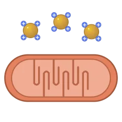 Mitochondria-targeted nanoparticle
 Cytosolic mRNA release and translation
 Intracellular trafficking of nanoparticles (endosome to lysosome)
Fate in the body 10
 EPR effect (nanoparticles leaking from tumour vessels)
 Normal vs tumour blood vessel (tight vs leaky)
 Active targeting of tumour cells by ligand-bearing nanoparticles
 Nanoparticle crossing the blood–brain barrier (receptor-mediated transcytosis)
 Renal clearance size cut-off (small particles filtered)
 Liver clearance by Kupffer cells
 Tumour penetration depth by particle size
 Mucus-penetrating vs mucoadhesive nanoparticles
 Inhaled nanoparticles reaching the alveoli
 Nanoparticles penetrating skin (via hair follicle)
Nanomedicine and theranostics 6
 Photothermal therapy of a tumour (NIR laser and gold nanoparticles)
 Photodynamic therapy (light, photosensitiser and singlet oxygen)
 Nanoparticle vaccine delivered to a dendritic cell
 Nanoparticle delivering CRISPR–Cas9 (ribonucleoprotein)
 Photoacoustic imaging with nanoparticles
 Artificial antigen-presenting nanoparticle activating a T cell
Making nanoparticles 15
 Microfluidic nanoparticle synthesis (herringbone mixer)
 Hydrodynamic flow focusing (microfluidic)
 Nanoprecipitation (organic solution dripped into stirred water)
 Double emulsion (water-in-oil-in-water) for particle making
 Self-assembly of amphiphiles into nanoparticles
 Seed-mediated growth of gold nanorods
 Citrate reduction synthesis of gold nanoparticles (boiling flask)
 Laser ablation in liquid (top-down synthesis)
 Thin-film hydration (lipid film to vesicles)
 Extrusion through a membrane (sizing vesicles)
 Probe sonication (breaking down particles)
 Top-down vs bottom-up nanofabrication
 Core–shell electrospun nanofibre (coaxial, drug in the core)
 Vertically aligned carbon nanotube forest (CVD growth)
 Nanoimprint lithography (stamp pressing a pattern)
Nanostructures and nanodevices 11
 Nanoneedle array delivering into a cell
 Bactericidal nanopillars (bacterium ruptured on a nanostructured surface)
 Track-etched nanoporous membrane
 Solid-state nanopore DNA sensing (with current trace)
 Nanowire field-effect transistor biosensor
 Nanocantilever sensor (bends when molecules bind)
 AFM tip imaging nanoparticles
 SERS substrate (plasmonic hot spots and Raman spectrum)
 Vertically aligned nanotube array (anodised titania)
 Nanoparticle superlattice (ordered array)
 Polymer nanocomposite (nanoparticles dispersed in a matrix)
Nanomotors and nanorobots 8
 Magnetic helical nanoswimmer (corkscrew nanorobot)
 Catalytic Janus nanomotor (bubble propulsion)
 Tubular microjet (rolled-up tube with a bubble tail)
 Enzyme-powered nanomotor (urease, fuel to propulsion)
 Light-driven nanomotor
 Ultrasound-propelled nanowire motor
 Biohybrid microrobot (bacterium pushing a cargo particle)
 Nanorobots travelling in a blood vessel
Characterisation 11
 Dynamic light scattering setup (laser, cuvette, detector)
 Monodisperse vs polydisperse nanoparticles
 AFM height profile of graphene quantum dots (1–2 nm)
 UV–vis absorption of carbon dots (π–π* and n–π* bands)
 Excitation-independent emission (overlapping normalised spectra)
 XPS survey of nitrogen-doped carbon dots (C, N, O peaks)
 XRD of carbon dots (broad (002) peak near 25°)
 Zeta potential distribution of carbon dots (negative surface)
 Fluorescence lifetime decay of carbon dots (TCSPC)
 Fluorescence spectrometer light path (emission at 90°)
 Elemental maps of nitrogen-doped carbon dots (C, N, O)
Carbon dot types 15
 Carbon quantum dot with a graphitic core (0.21 nm fringes)
 Carbonised polymer dot (polymer shell on a carbonised core)
 Few-layer graphene quantum dot with edge groups (side view)
 Nitrogen-doped carbon dot (N atoms in the lattice)
 Gadolinium-doped carbon dot (Gd³⁺ sites with bound water)
 Chiral carbon dots (L and D mirror pair)
 Red/NIR-emitting carbon dot (large conjugated domain)
 Carbon dot structure (sp² core and sp³ shell)
 Carbon dot surface groups (carboxyl, hydroxyl, amino)
 Molecular fluorophores (IPCA) bound to a carbon dot
 PEG-passivated carbon dot
 Carbon dot family (CQD, GQD and CPD)
 Carbon dot beside an antibody and a gold nanoparticle (to scale)
 Carbon dots embedded in a silica shell (CD@SiO₂)
 Carbon dot in water (hydration shell and counter-ions)
Bottom-up synthesis 11
 Hydrothermal synthesis in a Teflon-lined autoclave
 Hydrothermal reactor parts (steel jacket, Teflon liner, cap)
 Microwave synthesis of carbon dots (domestic oven)
 Reaction colour from clear to dark brown (carbonisation)
 Pyrolysis of citric acid (melt in a heating mantle)
 Bottom-up formation of a carbon dot (molecules, polymer, carbonised core)
 Carbon dots from orange peel (biomass precursor)
 Carbon dots from spent coffee grounds (carbonised in a furnace)
 Ultrasonic synthesis of carbon dots (sonochemical)
 Template synthesis of carbon dots in mesoporous silica
 Continuous-flow synthesis of carbon dots (heated coil reactor)
Top-down synthesis 5
 Carbon dots from candle soot
 Laser ablation of a graphite suspension (focused pulses)
 Arc-discharge soot between graphite electrodes
 Hydrothermal cutting of graphene oxide into dots
 Top-down and bottom-up routes to carbon dots
Purification and processing 6
 Dialysis of carbon dots (1 kDa membrane)
 Centrifuging carbon dots (dark pellet, clear supernatant)
 Silica column separating carbon dots by emission colour
 Carbon dot powder redissolving in water
 Stirred ultrafiltration cell for carbon dots
 Precipitating carbon dots with acetone
Optical properties 11
 Carbon dot solution in daylight and under UV light
 Where carbon dot emission comes from (core, surface and molecular states)
 Aggregation-caused quenching of carbon dots
 Solid-state fluorescent carbon dot powder under UV
 Room-temperature phosphorescence afterglow (UV on, UV off)
 Two-photon excitation of carbon dots (focal-spot emission)
 Photostability of carbon dots compared with an organic dye
 Photoinduced electron transfer from a carbon dot
 Electrochemiluminescence of carbon dots at an electrode
 Emission red-shifting with surface oxidation
 Carbon dot fluorescence and phosphorescence (energy diagram)
Sensing 12
 Fe³⁺ quenching a carbon dot (turn-off sensing)
 Turn-on sensing (quencher pulled off, glow restored)
 Fluorescence quenching titration (emission falling as ions are added)
 Selectivity of a carbon dot sensor across metal ions
 Carbon dot test paper for Hg²⁺ under UV
 pH-responsive carbon dot emission
 Carbon dot nanothermometer (emission falling with temperature)
 Ratiometric carbon dot probe (reference and responsive peaks)
 Smartphone reading a carbon dot test under UV
 Carbon dot–modified electrode for electrochemical sensing
 Peroxidase-like carbon dots turning TMB blue
 Cells with carbon dots before and after adding Fe³⁺
Bioimaging 11
 Cells labelled with carbon dots (glowing cytoplasm, dark nuclei)
 Nucleolus-targeting carbon dots
 Mitochondria-targeting carbon dots
 Plasma membrane–targeting carbon dots (bright cell outlines)
 Same cells imaged in three colour channels
 Renal clearance of carbon dots through the glomerular filter
 Ex vivo organ fluorescence after carbon dot injection
 Zebrafish larva imaged with carbon dots
 Bacteria labelled with carbon dots
 Folate-targeted carbon dots binding a cancer cell
 Cell viability with carbon dots (MTT assay)
Therapy and delivery 12
 Doxorubicin loaded on a carbon dot (π–π stacking)
 Doxorubicin released from carbon dots in an acidic endosome
 PEI–carbon dots carrying plasmid DNA (gene delivery)
 Photodynamic therapy with carbon dots (singlet oxygen)
 Photothermal therapy of a tumour with NIR-absorbing carbon dots
 Carbon dots damaging a bacterial membrane
 Carbon dots penetrating a bacterial biofilm
 Carbon dots blocking virus attachment
 Theranostic carbon dot (imaging, targeting and drug in one)
 Carbon dots inhibiting amyloid fibril growth
 Antioxidant carbon dots scavenging reactive oxygen species
 Carbon dot hydrogel dressing on an infected wound
Devices and other uses 9
 Carbon dot LED device stack (electroluminescence)
 Flexible light-emitting carbon dot film
 Luminescent solar concentrator with carbon dots
 Carbon dot–TiO₂ photocatalyst breaking down a dye
 Photocatalytic hydrogen evolution with carbon dots
 Carbon dots taken up by plant roots (UV view)
 Light-conversion greenhouse film with carbon dots
 Fluorescent carbon dot hydrogel
 Carbon dots boosting plant growth (treated and control)
2D nanomaterials 12
 Black phosphorus nanosheets (puckered phosphorene layers)
 MoS₂ layers (S–Mo–S sandwiches, side view)
 CVD-grown MoS₂ triangular domains on a wafer
 Hexagonal boron nitride nanosheet (white graphene)
 MXene made from a MAX phase (A layers etched out)
 Liquid-phase exfoliation of a layered crystal
 Van der Waals heterostructure (stacked 2D crystals)
 Crumpled graphene ball (paper-ball-like particle)
 Nanoscroll (sheet rolled into a spiral tube)
 Holey graphitic carbon nitride nanosheet (g-C₃N₄)
 Black phosphorus degrading in air and water
 Scotch-tape exfoliation (micromechanical cleavage)
Nanocrystal shapes 13
 Platinum–nickel octahedral nanocrystal (Pt₃Ni)
 Platinum–nickel nanoframe (hollow, edges only)
 Concave gold nanocube (high-index facets)
 Zinc oxide nanobelts (flat twisting ribbons)
 Zinc oxide nanospring (nanohelix)
 Gold–iron oxide dumbbell nanoparticle (heterodimer)
 Nanoporous gold (dealloyed sponge)
 Silicon nanowires grown by VLS (gold droplet tips)
 Calcium carbonate (vaterite) microsphere
 Striped metal nanowires (nanobarcodes, Au/Ag/Ni)
 Liquid-metal nanodroplets (gallium alloy, oxide skin)
 Oleic-acid-capped nanocrystal (hydrophobic ligand shell)
 Iron oxide nanoflower (multicore, oriented grains)
Frameworks and porous crystals 7
 UiO-66 octahedral crystal (zirconium MOF)
 Breathing MOF (MIL-53 pores open and close)
 Enzyme encapsulated in ZIF-8 (biomimetic mineralisation)
 MOF-on-MOF core–shell crystal
 Zeolite crystal (ZSM-5 coffin habit, channel openings)
 Surface-mounted MOF film grown layer by layer (SURMOF)
 ZIF-8 dissolving in acid (cargo release)
Protein and peptide assemblies 9
 Encapsulin nanocompartment (cargo inside a protein shell)
 Vault ribonucleoprotein particle (barrel)
 Cyclic peptide nanotube (stacked rings)
 Self-assembling peptide nanofibre mesh (RADA16 hydrogel)
 Elastin-like polypeptide coacervation (heat-triggered)
 Isolated magnetosome chain (membrane-wrapped magnetite)
 Cellulose nanocrystals (rod-like CNCs, TEM)
 HDL-mimetic nanoparticle (gold core, apoA-I belts)
 Ferritin disassembly and reassembly (drug loading)
Cell-derived carriers 12
 Red-cell-membrane nanosponge soaking up toxins
 Platelet-membrane nanoparticles binding an injured vessel
 Neutrophil-membrane nanoparticle soaking up cytokines
 Membrane coating by co-extrusion
 Cancer-cell-membrane nanoparticles homing to their source cells
 Hybrid-membrane coating (red cell and platelet membranes fused)
 Nanoparticles hitchhiking on red blood cells
 Cellular backpack on a macrophage (adhered polymer disc)
 T cell carrying surface-anchored cytokine nanogels
 Engineered exosome displaying a targeting peptide (Lamp2b fusion)
 Bacterial minicell (anucleate, drug-loaded)
 Yeast glucan particle (hollow β-glucan shell)
DNA assemblies 10
 DNA bricks assembling into a 3D shape
 Wireframe DNA polyhedron (icosahedral mesh)
 DNA nanoflower grown by rolling-circle amplification
 DNA hydrogel (cross-linked X-shaped building blocks)
 DNA tweezers (opened and closed by fuel strands)
 DNA origami nanoruler (super-resolution calibration)
 Gold nanoparticle helix on DNA origami (plasmonic chirality)
 Nanoflare (target mRNA releases a fluorescent flare)
 Tetrahedral DNA probes on a gold electrode (electrochemical sensor)
 DNA origami presenting ligands at set spacings
Corona and colloids 10
 Protein corona masking targeting ligands
 Opsonised nanoparticle recognised by a macrophage
 Protein unfolding on a nanoparticle surface
 Eco-corona on a nanoparticle in natural water
 Isolating the protein corona (spin, wash, elute, gel)
 Accelerated blood clearance of a second PEGylated dose
 Steric vs electrostatic stabilisation of colloids
 Particle sedimentation onto cells in a well (delivered dose)
 Zinc oxide nanoparticle dissolving into ions
 Agglomerate vs aggregate (loosely held vs fused)
Uptake and escape 11
 Endosomal escape by membrane fusion (fusogenic lipids)
 Endosomal escape through peptide pores
 Photochemical internalisation (light ruptures endosomes)
 Ionisable lipids forming a non-bilayer phase with the endosome membrane
 Galectin puncta marking damaged endosomes
 Nanoparticles captured by autophagy
 Membrane wrapping depends on particle size
 Particle shape and uptake (rods entering tip-first)
 Soft vs stiff particles at the cell membrane
 Nanoparticles passed between cells along a tunnelling nanotube
 Blocking endocytosis routes with inhibitors
Delivery in the body 13
 Active trans-endothelial transport into a tumour
 Splenic filtration of rigid particles (interendothelial slits)
 Hepatobiliary clearance (hepatocyte to bile)
 Lymphatic drainage of nanoparticles to a lymph node
 Oral nanoparticles crossing the gut via M cells
 Size-shrinkable nanoparticle (large in blood, small in tumour)
 Charge-reversal nanoparticle in acidic tumour tissue
 Neutrophils carrying nanoparticles across a vessel wall
 Dense collagen stroma blocking nanoparticle spread
 High interstitial fluid pressure pushing particles back
 ApoE corona steering lipid nanoparticles to hepatocytes
 Focused ultrasound with microbubbles opening the blood–brain barrier
 Nanoparticles at the placental barrier
Nanotherapies and imaging 10
 Peroxidase-like iron oxide nanozyme turning TMB blue
 Manganese dioxide nanosheets making oxygen in a hypoxic tumour
 Chemodynamic therapy (Fenton reaction, hydroxyl radicals)
 Glucose oxidase nanoreactor (tumour starvation)
 Sonodynamic therapy (ultrasound activates a sonosensitiser)
 Clot-targeted nanoparticles releasing tPA (thrombolysis)
 Nanoparticles penetrating a bacterial biofilm
 Iron-based nanoparticles driving ferroptosis (lipid peroxidation)
 Radiolabelled nanoparticle for PET imaging
 Trojan particles for inhalation (nanoparticles in porous microparticles)
Plasmonics and sensing 10
 SERS tag (gold core, Raman reporter, silica shell)
 Plasmonic bowtie nanoantenna (gap hot spot)
 Plasmonic nanohole array (extraordinary transmission)
 LSPR sensing (peak shift when molecules bind)
 Plasmon ruler (colour shifts with gap distance)
 Tip-enhanced Raman spectroscopy (TERS)
 Metal-enhanced fluorescence vs quenching (distance from gold)
 Bio-barcode assay (gold probes release barcode DNA)
 Magnetic relaxation switch (particles cluster when target binds)
 Graphene oxide quenching dye-labelled probes (turn-on sensor)
Micro- and nanorobots 6
 Gold–platinum nanorod motor (self-electrophoresis)
 Algae-driven microrobot (microalga carrying cargo)
 Electromagnetic coil system steering a microrobot
 Magnetic microrobot swarm (rotating field)
 Magnetic microroller climbing a vessel wall
 Magnesium micromotor propelled by stomach acid (H₂ bubbles)
Catalysis and energy 8
 Single-atom catalyst (isolated metal atoms on a support)
 Platinum nanoparticles on carbon black (fuel-cell catalyst)
 Plasmonic hot-electron transfer (gold on titania)
 Silicon nanoparticle anode swelling on lithiation
 Dye-sensitised solar cell (TiO₂ nanoparticle film)
 Photocatalytic water splitting on a cocatalyst-loaded particle
 Quantum-dot light-emitting diode (QLED layer stack)
 Plasmonic solar steam generation (floating absorber film)
Nanofabrication 15
 Photolithography (UV through a photomask)
 Electron-beam lithography (writing in resist)
 Dip-pen nanolithography
 Block copolymer self-assembly patterns (lamellae and dots)
 Nanosphere lithography (triangle array from a colloidal mask)
 Layer-by-layer assembly by sequential dipping
 Atomic layer deposition (ALD) cycle
 Graphene grown by CVD on copper foil
 Sputter coating (argon plasma, gold target)
 Reactive ion etching (anisotropic plasma etch)
 Lift-off patterning of metal
 Template synthesis of nanowires in anodic alumina
 Glancing-angle deposition (nanohelices)
 Langmuir–Blodgett transfer (trough and dipper)
 Metal-assisted chemical etching of silicon nanowires
Colloidal and aerosol synthesis 7
 Hot-injection synthesis of nanocrystals (three-neck flask)
 Galvanic replacement (silver cube to hollow gold–silver box)
 Nanoscale Kirkendall effect (solid particle to hollow shell)
 Phase transfer of nanocrystals by ligand exchange
 Reverse microemulsion synthesis (water droplets as nanoreactors)
 Co-precipitation of iron oxide nanoparticles (Fe²⁺/Fe³⁺ plus base)
 Flame spray pyrolysis